Why Eye Care Exams Are Critical for Detecting Serious Conditions Early
Routine health check-ups typically prioritize cardiovascular screening, metabolic blood panels, and dental maintenance, while comprehensive vision evaluations are frequently pushed aside. Many individuals mistakenly believe that an eye care exam is only necessary when their physical prescription updates or when they experience sudden, noticeable blurring. In reality, the human eye serves as a unique physiological window through which specialized clinicians can directly observe active vascular and neurological structures in real time.
This comprehensive article explores the vital role that routine ocular testing plays in safeguarding your visual acuity and identifying systemic health conditions long before external symptoms emerge. By examining the structural, biological, and preventive elements of these evaluations, property managers of their own health can implement highly effective wellness strategies. Partnering with a dedicated clinic ensures that minor optical shifts are managed early, protecting both your structural vision and your long-term internal health.
The Crucial Need for Early Clinical Intervention
Regular diagnostic checks are the primary line of defense against progressive ocular diseases that intentionally develop without early warnings or painful symptoms. Chronic conditions like open-angle glaucoma, age-related macular degeneration, and premature cataracts can quietly destroy delicate retinal cells over several years. Utilizing an eye exam allows optometrists to analyze structural micro-shifts before irreversible peripheral vision loss occurs, drastically reducing the necessity for invasive surgical interventions or intensive pharmaceutical treatments down the line.
In many scenarios, a patient remains entirely unaware of their visual degradation because the brain automatically compensates for focal blind spots. For instance, advanced glaucoma causes slow, permanent damage to the optic nerve matrix without triggering any initial physical pain.
Catching these structural changes early is the only definitive method to alter the disease trajectory. According to the 2023 National Health Interview Survey, 51.9 million American adults have some difficulty seeing, including 3.7 million with severe vision loss, with women representing 57% of affected adults. This vast demographic burden underscores the critical nature of scheduling routine, preventative clinical evaluations to preserve independent living.
A strategic proactive approach to ocular wellness involves blending regular clinical diagnostic testing with targeted lifestyle management. During an eye care exam, specialists provide customized guidance regarding UV-blocking lens selections, nutritional optimization, and blue-light screen filters to reduce chronic digital strain. These custom recommendations are highly beneficial for managing underlying metabolic conditions that directly degrade the micro-vasculature of the retina over time.
Ocular testing frequencies must be systematically tailored to an individual’s age, ancestral risk profile, and systemic health status. Medical guidelines generally advise that healthy adults aged 18 to 64 undergo a comprehensive check every twenty-four months, while seniors over 65 should commit to an annual schedule due to increased disease risks.
Pediatric patients require initial structural screenings at six months, three years, and right before entering primary school. Catching structural anomalies like amblyopia early prevents long-term academic developmental delays, ensuring children possess the clear visual focus necessary to thrive.
Diagnostic Tests and Ocular Architecture
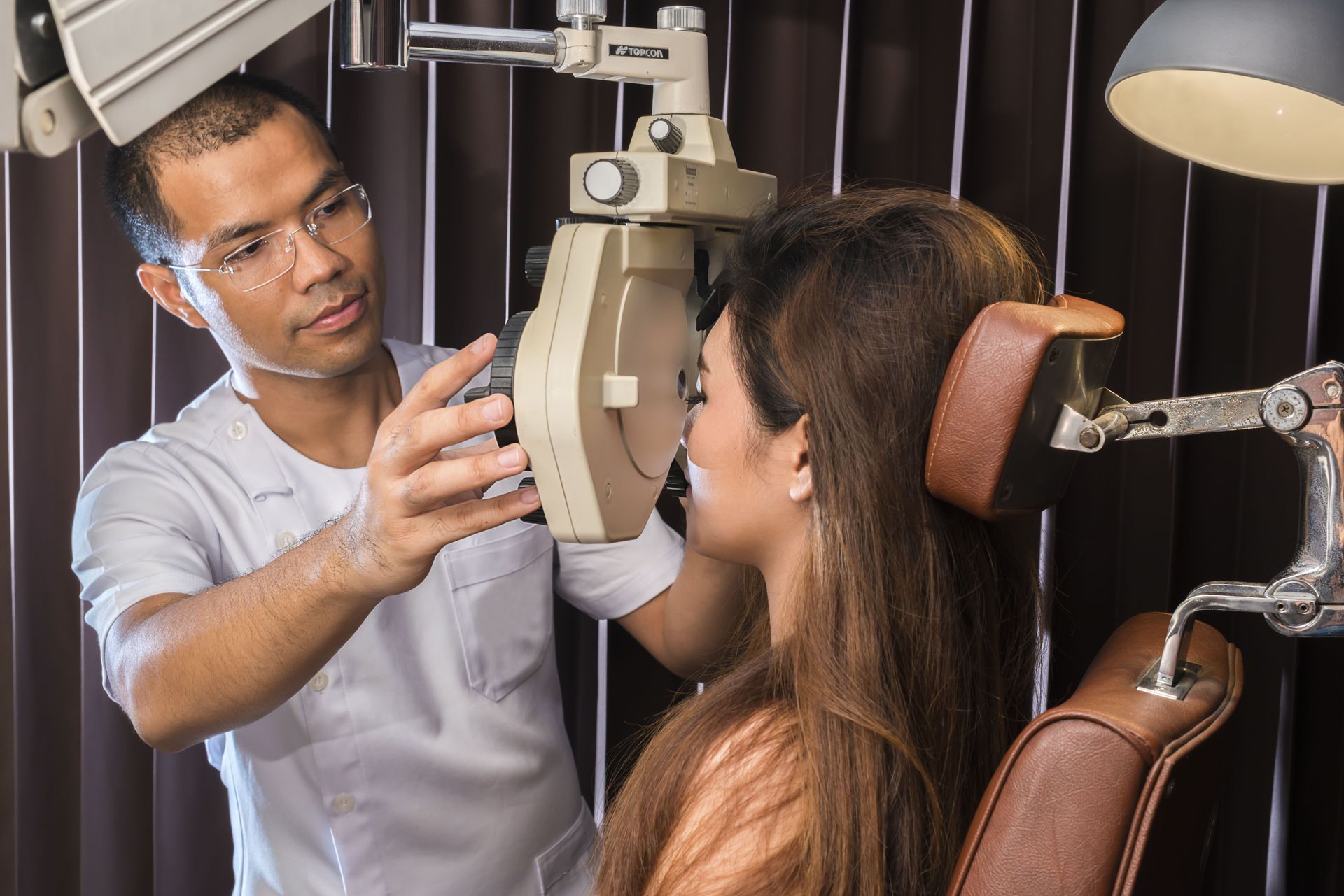
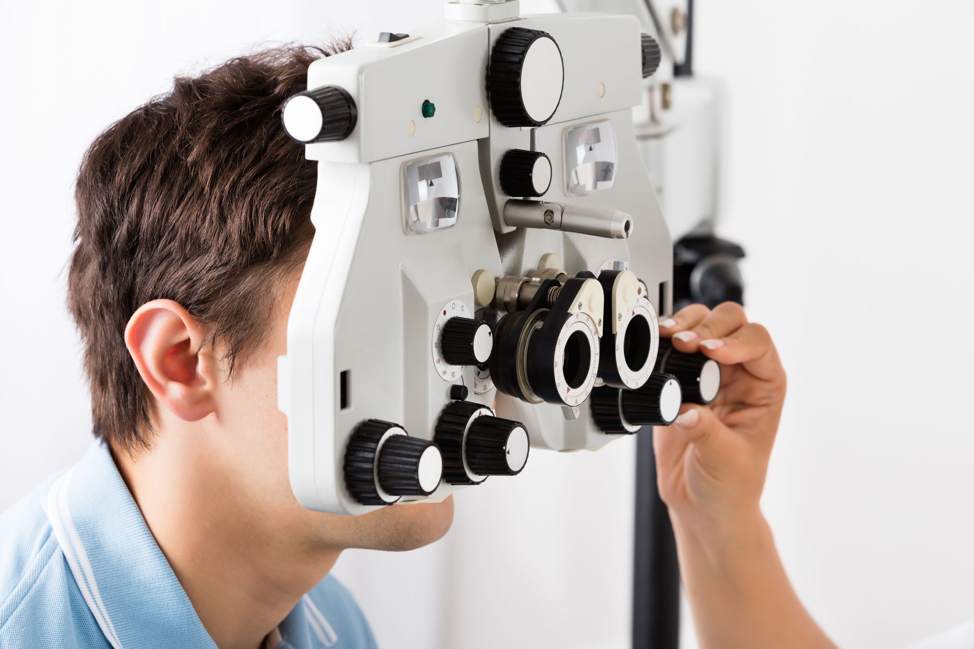
A comprehensive eye care exam utilizes an array of specialized technical tests designed to map the external and internal topography of the eye. Beyond standard letter charts that measure visual acuity, clinicians employ precision refraction testing to calculate exact corrective lens requirements. A specialized slit-lamp examination utilizes a high-magnification biomicroscope to inspect the anterior structures, including the cornea, iris, and crystalline lens, identifying microscopic signs of cataracts or localized inflammation.
To evaluate glaucoma risks, clinicians use non-contact or applanation tonometry to measure intraocular pressure within the aqueous humor. Advanced clinics enhance this diagnostic suite with Optical Coherence Tomography, which takes cross-sectional, high-resolution scans of the macular layers. These digital scans map out microscopic fluid leaks and cellular thinning long before they cause visual distortion, serving as an invaluable baseline for preventive health management.
Identifying Systemic Diseases Through Ocular Tracking
The microscopic blood vessels supplying the retina are highly sensitive to chronic elevations in blood glucose levels. During a comprehensive eye exam, a clinician can identify early structural changes associated with diabetic retinopathy, including micro-aneurysms, localized hemorrhages, and hard lipid exudates. Identifying these subtle vascular abnormalities allows primary care physicians to adjust metabolic management programs quickly, preventing proliferative blindness.
Chronic high blood pressure forces the delicate arterial networks of the eye to constrict and thicken to protect retinal tissues. A dilated eye care exam allows specialists to view these vascular shifts, mapping out copper-wiring arteriole changes, focal ischemia, and retinal swelling. These ocular markers provide direct insight into the patient’s systemic cardiovascular strain, serving as an early warning for potential strokes or heart attacks.
Systemic autoimmune disorders, such as rheumatoid arthritis, consistently cause inflammatory damage to ocular tissues. During a thorough eye exam, a specialist can identify severe dry eye syndrome, episcleritis, or deep intraocular inflammation. Tracking these localized flare-ups assists rheumatologists in fine-tuning immunosuppressive therapies, effectively protecting the eye while reducing systemic joint pain.
Elevated blood lipids can display clear structural indicators on the exterior and interior structures of the eye. Younger patients undergoing an eye care exam may display corneal arcus—a distinct gray-white lipid ring circling the outer edge of the cornea—or yellowish cholesterol plaques on the eyelids. Internally, these fatty deposits can trigger catastrophic retinal artery occlusions, demanding immediate cardiovascular intervention.
Autoimmune thyroid conditions, particularly Graves' disease, lead to severe inflammation and swelling within the orbital muscles and fat pockets behind the eye. Ocular check-ups monitor for characteristic signs like upper eyelid retraction, restricted muscle tracking, and exophthalmos. Regular clinical monitoring prevents optic nerve compression and corneal exposure ulcerations, protecting comfort and visual acuity throughout the endocrine treatment cycle.
Optic Nerve Integrity and Neurological Health
The optic nerve functions as a specialized extension of the central nervous system, connecting the retina directly to the visual cortex of the brain. Evaluating the optic disc during an eye care exam provides critical insights into a patient's broader neurological condition. For example, bilateral swelling of the optic disc directly signals dangerously elevated intracranial pressure, necessitating emergency neurological imaging to rule out brain tumors or cerebrospinal fluid blockages.
Furthermore, localized inflammation of the nerve fibers, known as optic neuritis, often presents as a primary clinical warning sign of demyelinating diseases like Multiple Sclerosis. By tracking changes in the nerve fiber layer and mapping peripheral blind spots through automated visual field testing, eye care professionals act as crucial members of multidisciplinary neurological teams, protecting brain health and preserving sight.
Prioritizing a comprehensive eye care exam is a fundamental step toward building long-term vision protection and robust systemic health management. These deep clinical diagnostics provide invaluable opportunities to identify sight-threatening conditions, metabolic complications, cardiovascular strain, and complex neurological shifts long before they affect your daily quality of life. By committing to an age-appropriate examination schedule, individuals can easily insulate their visual performance from irreversible degradation, ensuring long-term independence and lasting wellness.
When managing your family’s optical health, relying on quick online vision screeners or uncertified over-the-counter reading lenses can leave silent, blind-spot diseases completely undetected until severe structural damage has occurred. Because early stages of sight-threatening conditions like glaucoma or diabetic retinopathy rarely alter your initial glasses prescription, a simple focal test cannot substitute for an in-depth biological mapping of the retina. True preventative health requires advanced digital diagnostic imaging, meticulous intraocular pressure tracking, and expert clinical evaluations from a board-certified specialist. To safeguard your vision with cutting-edge diagnostics, customized lens prescriptions, and elite patient care, consider scheduling your next eye care exam with the trusted team at Optical City.


Share On: